
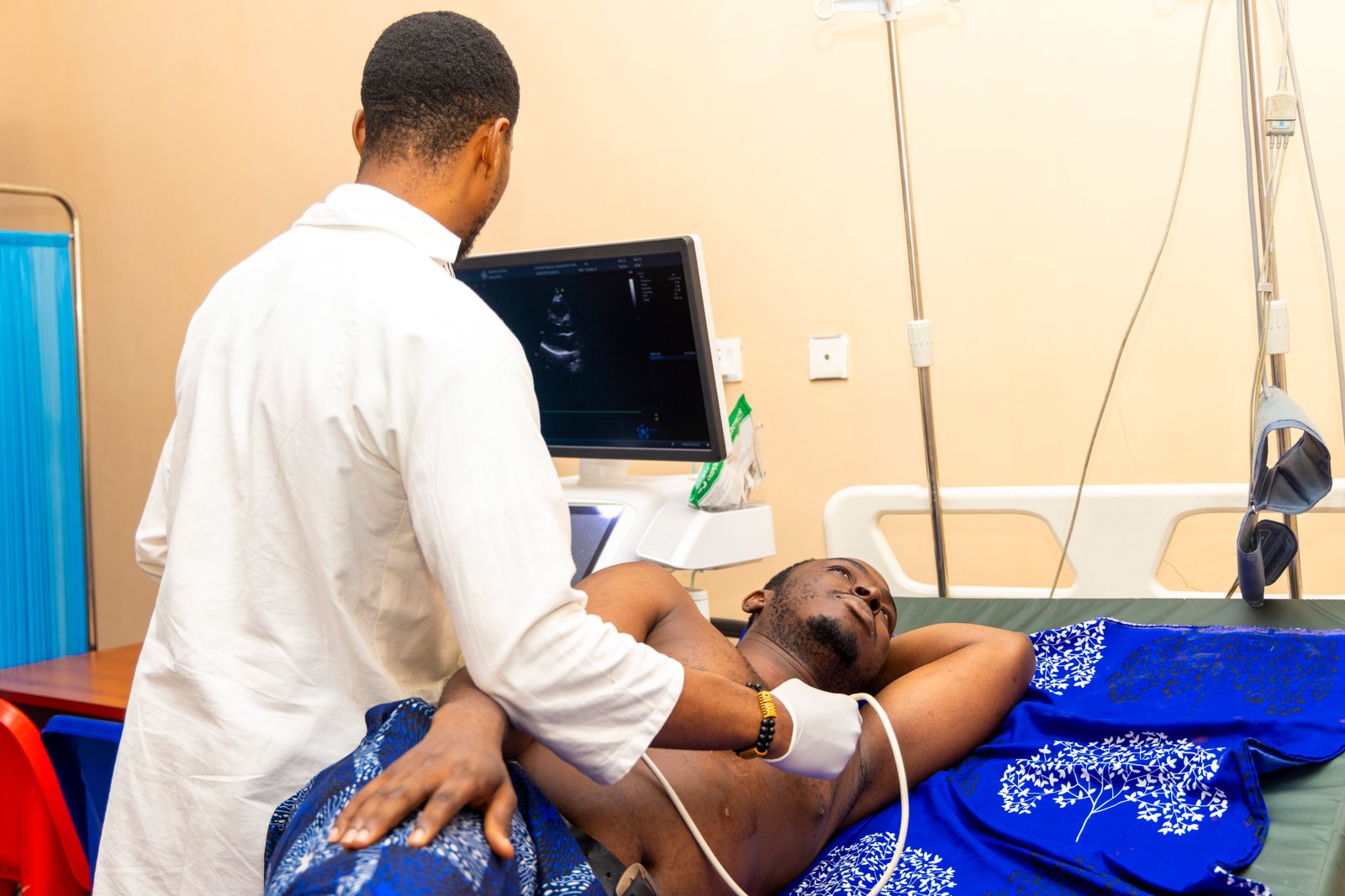
Echocardiography Services in Owerri
Echocardiogram Testing for Heart Structure and Function
The heart works continuously to pump blood throughout the body, and understanding how it is functioning can be an important part of diagnosing and monitoring many heart conditions.
At LifeWay Diagnostics, we provide professional echocardiography services in Owerri using ultrasound-based imaging to examine the heart’s structure, movement and function.
An echocardiogram can help healthcare professionals assess the heart chambers, heart valves, pumping function and blood flow, providing valuable information alongside other clinical examinations and cardiac tests.
Whether you have been referred for an echocardiogram because of specific symptoms, are being monitored for an existing heart condition, or require cardiac assessment as part of your medical evaluation, our team is available to support your diagnostic needs.
Need an echocardiogram in Owerri? Contact LifeWay Diagnostics to make an enquiry or confirm your appointment.
What Is an Echocardiogram?
An echocardiogram, commonly called an echo, is an ultrasound examination of the heart.
Instead of using X-rays, echocardiography uses high-frequency sound waves to create moving images of the heart. These images allow healthcare professionals to examine how the heart is structured and how it moves as it beats.
Depending on the examination and clinical request, echocardiography can provide information about:
The size and shape of the heart chambers
Movement of the heart walls
How well the heart is pumping
The appearance and movement of the heart valves
Blood flow through the heart
The relationship between different parts of the heart
Certain abnormalities affecting the structure or function of the heart
An echocardiogram is often interpreted together with your symptoms, medical history, physical examination and other investigations.
Why Might You Need an Echocardiogram?
A doctor may request an echocardiogram when they need more information about the structure or function of your heart.
It may be recommended for people experiencing symptoms such as:
Chest discomfort or pain
Shortness of breath
Unusual tiredness
Dizziness or fainting
Heart palpitations
Swelling of the legs or feet
Reduced exercise tolerance
Symptoms that may be associated with heart disease
An echocardiogram may also be requested when a healthcare professional detects a heart murmur or wants to investigate a suspected cardiac abnormality.
For people who already have a diagnosed heart condition, echocardiography may also be used as part of ongoing monitoring.
An echocardiogram does not by itself determine the cause of every heart-related symptom. Your doctor or healthcare professional will consider the results alongside your overall clinical picture.
What Can an Echocardiogram Show?
Echocardiography provides detailed information about the heart and can help healthcare professionals evaluate several important features.
Heart Chambers
The heart has four chambers that work together to circulate blood through the lungs and the rest of the body.
An echocardiogram can provide information about the size and appearance of these chambers and how they are functioning.
Heart Valves
The heart contains valves that help blood move in the correct direction.
Echocardiography can assess the appearance and movement of the valves and can provide information about blood flow across them.
This can be useful when investigating conditions involving valve narrowing or leakage.
Heart Pumping Function
The heart needs to contract effectively to pump blood around the body.
An echocardiogram can help assess how well the heart muscle is moving and pumping.
Blood Flow
With Doppler ultrasound techniques, echocardiography can assess blood flow through the heart and across the valves.
This can provide additional information when evaluating certain cardiac conditions.
Heart Muscle Movement
An echocardiogram can show how different sections of the heart muscle move during the cardiac cycle.
Changes in heart-wall movement may provide important information that doctors can consider alongside symptoms and other investigations.
What Happens During an Echocardiogram?
An echocardiogram is generally a straightforward and non-invasive examination.
When you arrive at LifeWay Diagnostics, you may be asked to provide your relevant clinical information or referral details.
During the examination:
1. You will be positioned for the scan
You will usually lie on an examination bed, often positioned partly on your side so the heart can be viewed clearly.
2. Electrodes may be placed on your chest
Small monitoring electrodes may be used to help record the timing of your heartbeat during the examination.
3. Ultrasound gel is applied
A small amount of ultrasound gel is placed on the chest. The gel helps the ultrasound probe make good contact with the skin.
4. The sonographer or healthcare professional moves the probe
A handheld ultrasound probe is gently placed against different areas of your chest.
You may feel some pressure as the probe is moved to obtain different views of the heart.
5. Images are recorded
The ultrasound system produces real-time images of the heart while it is beating.
Different views may be obtained to assess the heart chambers, valves, muscle movement and blood flow.
6. The examination is completed
Once the necessary images have been obtained, the equipment and gel are removed and you can normally continue with your day unless your healthcare professional advises otherwise.
Is an Echocardiogram Painful?
An echocardiogram is generally not painful.
You may feel mild pressure when the ultrasound probe is pressed against certain areas of your chest to obtain a clearer image.
Some people may find maintaining a particular position or taking certain breaths slightly uncomfortable, but the examination is generally well tolerated.
If you experience significant discomfort during the procedure, let the healthcare professional performing the examination know.
How Long Does an Echocardiogram Take?
The duration can vary depending on the type of examination, the views required and the clinical reason for the test.
A standard echocardiogram may take approximately 30 to 60 minutes, although the exact duration can vary.
Your healthcare professional can provide more specific information about your appointment.
Do I Need to Prepare for an Echocardiogram?
For a standard echocardiogram, extensive preparation is usually not required.
However, preparation requirements can depend on the specific examination and your clinical circumstances.
Before your appointment:
Follow any instructions provided by LifeWay Diagnostics or your referring doctor.
Bring your referral or request form if applicable.
Bring relevant previous cardiac investigation reports if available.
Tell the healthcare professional about any relevant medical conditions or concerns.
Wear clothing that allows appropriate access to the chest area.
If you have been given specific preparation instructions, follow those instructions rather than relying on general information.
Is Echocardiography Safe?
Echocardiography uses ultrasound waves rather than ionizing radiation.
Standard echocardiography is considered a non-invasive diagnostic imaging examination and is widely used for assessing the heart.
If you are pregnant, breastfeeding or have concerns about the safety of a particular examination, discuss this with your healthcare professional before the procedure.
Your doctor or diagnostic team can advise whether the requested examination is appropriate for your individual circumstances.
Echocardiography and Pregnancy
Because standard echocardiography uses ultrasound rather than X-rays, ultrasound-based imaging is commonly used in medical care, including during pregnancy when clinically indicated.
If you are pregnant or think you may be pregnant, inform the healthcare professional before your examination.
Your doctor will determine whether an echocardiogram is appropriate based on your medical needs.
Echocardiography vs ECG: What Is the Difference?
An ECG and an echocardiogram are both important cardiac investigations, but they examine different aspects of the heart.
ECG — Electrocardiography
An ECG records the electrical activity of the heart.
It can provide information about:
Heart rate
Heart rhythm
Electrical activity
Certain rhythm abnormalities
Some patterns that may suggest cardiac problems
Echocardiogram
An echocardiogram uses ultrasound to produce images of the heart.
It can provide information about:
Heart chambers
Heart valves
Heart muscle movement
Pumping function
Blood flow
In some situations, a doctor may request both an ECG and an echocardiogram because the tests provide different types of information.
Echocardiography vs Other Heart Tests
Different cardiac investigations answer different clinical questions.
An ECG focuses primarily on the heart’s electrical activity.
An echocardiogram provides ultrasound images of the heart’s structure and function.
A Holter monitor records heart rhythm over a longer period while you go about your normal activities.
Other investigations may be requested depending on your symptoms, medical history and clinical assessment.
Your doctor will determine which test or combination of tests is appropriate for your situation.
Who May Be Referred for an Echocardiogram?
Echocardiography may be requested for people with suspected or known cardiovascular conditions.
Your doctor may recommend an echocardiogram if there are concerns about:
Heart valve abnormalities
Heart chamber enlargement
Heart muscle function
Certain structural heart abnormalities
Abnormal heart sounds or murmurs
Symptoms that may have a cardiac cause
Changes in cardiac function requiring monitoring
It may also be used as part of an assessment when a healthcare professional wants more information about the heart after a clinical examination.
What Happens After the Echocardiogram?
After the examination, the images and findings are reviewed according to the diagnostic process.
Depending on the arrangement for your examination, a report may be provided to your referring doctor or made available to you according to the centre’s reporting procedure.
Your doctor will interpret the result in the context of:
Your symptoms
Medical history
Physical examination
Previous investigations
Other laboratory or imaging results
A finding on an echocardiogram does not necessarily provide a complete diagnosis on its own.
If further investigation or treatment is required, your doctor can advise you on the appropriate next steps.
When Should You Speak to a Doctor About Heart Symptoms?
Some symptoms should not be ignored.
If you experience new, severe or persistent chest pain, significant difficulty breathing, fainting or other potentially serious symptoms, seek urgent medical attention rather than waiting for a routine diagnostic appointment.
For non-emergency concerns, speak with a qualified healthcare professional who can assess your symptoms and determine whether an echocardiogram or another investigation is appropriate.
Echocardiography in Owerri at LifeWay Diagnostics
At LifeWay Diagnostics, our echocardiography service forms part of a broader range of diagnostic and clinical investigation services available in Owerri.
Our approach is focused on providing patients and referring healthcare professionals with reliable diagnostic information within a professional clinical environment.
Whether you have been referred by a doctor, are undergoing investigation for specific symptoms, or require follow-up for an existing cardiac concern, we can help you access the appropriate diagnostic service.
Why patients and referring professionals may choose LifeWay Diagnostics
Professional diagnostic environment
Access to cardiac and clinical diagnostic services
Modern diagnostic equipment
Patient-focused service
Convenient access to multiple diagnostic services
Support for doctor referrals and clinical investigations
Integration with other laboratory, imaging and physiology services
Our goal is to make diagnostic care more accessible to people in Owerri and surrounding communities while supporting healthcare professionals with useful diagnostic information.
Related Cardiac and Diagnostic Services
Echocardiography is part of a wider diagnostic pathway. Depending on your clinical needs, your healthcare professional may also request other investigations.
Explore related services at LifeWay Diagnostics:
ECG in Owerri
Electrocardiography records the electrical activity and rhythm of the heart.
Holter Monitoring
Holter monitoring can record heart rhythm over an extended period, which may be useful when symptoms do not occur during a short ECG.
Ambulatory Blood Pressure Monitoring
ABPM records blood pressure at intervals over an extended period to provide information about blood pressure patterns during normal daily activities.
Clinical Physiology
Explore LifeWay Diagnostics’ broader clinical physiology services, including cardiac, respiratory and neurological investigations.
Radiology & Medical Imaging
Access imaging services including MRI, CT, ultrasound, X-ray and mammography.
Laboratory Services
Laboratory investigations can provide additional information that may support the evaluation of a patient’s overall health.
Frequently Asked Questions About Echocardiography in Owerri
What is an echocardiogram?
An echocardiogram is an ultrasound examination used to create moving images of the heart. It can provide information about the heart chambers, valves, muscle movement, pumping function and blood flow.
Is an echocardiogram the same as an ECG?
No. An ECG records the electrical activity of the heart, while an echocardiogram uses ultrasound to produce images of the heart’s structure and function.
Is an echocardiogram painful?
An echocardiogram is generally not painful. You may experience some pressure from the ultrasound probe as it is placed against your chest.
How long does an echocardiogram take?
The examination commonly takes around 30 to 60 minutes, although the duration can vary depending on the examination and clinical requirements.
Do I need to fast before an echocardiogram?
Standard echocardiography usually does not require fasting. However, preparation can vary depending on the specific examination, so follow the instructions provided for your appointment.
Does an echocardiogram use radiation?
Standard echocardiography uses ultrasound rather than ionizing radiation.
Can pregnant women have an echocardiogram?
When clinically indicated, ultrasound-based examinations may be used during pregnancy. Inform your healthcare professional if you are pregnant so that your care can be appropriately planned.
What can an echocardiogram detect?
An echocardiogram can provide information about heart structure and function, including heart chambers, valves, heart muscle movement and blood flow. Your doctor interprets the findings alongside your symptoms and other clinical information.
Do I need a doctor’s referral for an echocardiogram?
This can depend on the clinical circumstances and the diagnostic centre’s appointment process. If you have been advised to have an echocardiogram, bring your doctor’s request or referral where applicable.
Where can I get an echocardiogram in Owerri?
LifeWay Diagnostics provides echocardiography and other diagnostic services in Owerri. Contact the centre to confirm availability, appointment requirements and any preparation instructions before visiting.
Book or Enquire About Echocardiography in Owerri
If your doctor has requested an echocardiogram or you have been advised to undergo cardiac assessment, LifeWay Diagnostics is available to assist.