
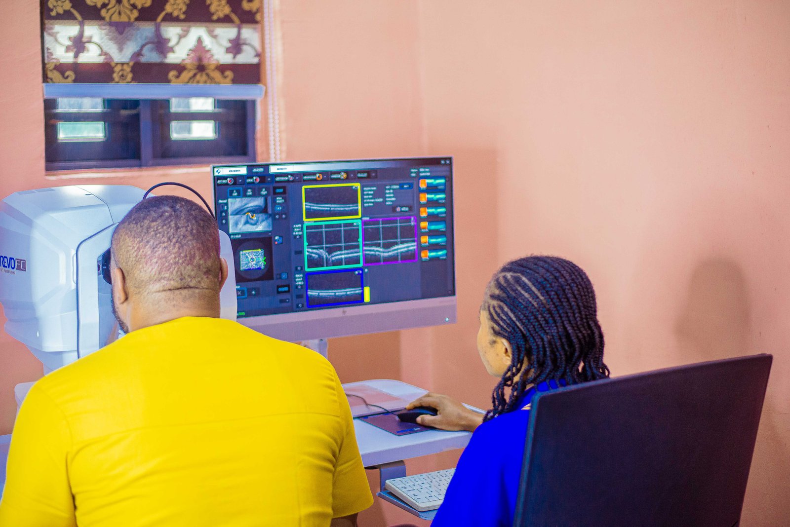
Optical Coherence Tomography (OCT) Services in Owerri
Advanced Eye Imaging for Detailed Examination of the Retina and Optic Nerve
When you need more than a routine eye examination, detailed imaging can help provide a clearer view of the structures inside the eye.
Optical Coherence Tomography (OCT) is a non-invasive imaging test that uses light to produce detailed, cross-sectional images of structures within the eye, particularly the retina and optic nerve.
At LifeWay Diagnostics in Owerri, OCT forms part of our Clinical Physiology and diagnostic services, providing detailed eye imaging that can support the clinical assessment and monitoring of various eye conditions.
Whether an OCT scan has been recommended by your eye doctor or you have been asked to undergo detailed retinal or optic nerve imaging, our team can guide you through the examination and explain what to expect.
What Is Optical Coherence Tomography?
Optical Coherence Tomography, commonly called OCT, is an advanced imaging technique used to obtain detailed images of the internal layers of the eye.
Unlike a conventional photograph of the eye, OCT can produce cross-sectional images that allow clinicians to examine individual retinal layers and other important structures.
The examination is generally:
Non-invasive
Painless
Quick to perform
Performed without touching the eye
Based on light rather than X-rays
OCT can provide valuable structural information that may not be visible during a standard examination alone.
What Does an OCT Scan Examine?
Depending on the type of examination and the clinical reason for the scan, OCT may be used to obtain detailed images of structures such as the:
Retina
The retina is the light-sensitive layer at the back of the eye responsible for receiving visual information.
OCT can provide detailed images of its different layers and help clinicians assess structural changes.
Macula
The macula is the central part of the retina responsible for detailed central vision.
OCT imaging can be particularly useful when a clinician needs to examine the macula for swelling, fluid, thinning or other structural changes.
Optic Nerve
The optic nerve carries visual information from the eye to the brain.
OCT can provide measurements and images that may assist in evaluating the optic nerve and monitoring changes over time.
Retinal Nerve Fibre Layer
OCT can also provide measurements of the retinal nerve fibre layer, which may be relevant when assessing or monitoring certain optic nerve conditions.
Why Might a Doctor Recommend an OCT Scan?
OCT may be recommended for different clinical reasons.
Your eye specialist may request an OCT when they need more detailed information about the retina, macula or optic nerve.
It may be used as part of the assessment or monitoring of conditions such as:
Glaucoma
Macular conditions
Diabetic eye disease
Retinal changes
Macular oedema
Age-related retinal changes
Optic nerve abnormalities
Other conditions affecting the retina or optic nerve
An OCT scan does not replace a complete eye examination. Instead, it provides additional structural information that can help a qualified eye-care professional understand what may be happening inside the eye.
OCT for Diabetic Eye Assessment
People living with diabetes can develop changes in the blood vessels and tissues of the retina.
Depending on the clinical situation, OCT may be used to examine retinal structure and identify changes that require further evaluation.
For patients undergoing ongoing eye care, OCT can also provide images that may be compared with previous examinations when clinically appropriate.
If you have diabetes, your doctor or eye specialist may recommend regular eye examinations even when your vision appears normal.
OCT and Glaucoma Assessment
Glaucoma can affect the optic nerve and may progress without obvious symptoms during its early stages.
OCT can provide detailed measurements and images of structures around the optic nerve and retinal nerve fibre layer.
These measurements may help an eye-care professional assess structural changes and monitor patients over time.
However, OCT alone does not diagnose glaucoma. It is normally considered alongside other clinical information and examinations, such as eye pressure measurements, visual field testing and assessment of the optic nerve.
OCT and Macular Conditions
The macula plays an important role in central vision.
Changes affecting the macula can sometimes lead to symptoms such as:
Blurred central vision
Distorted vision
Difficulty reading
Difficulty seeing fine details
Changes in the appearance of straight lines
A noticeable area of reduced vision
OCT can produce detailed cross-sectional images of the macula, allowing clinicians to examine its structure and identify changes that may require further investigation or treatment.
What Happens During an OCT Scan?
An OCT examination is generally straightforward and does not require instruments to touch the surface of your eye.
Step 1: Patient Information
You may be asked about your symptoms, medical history, eye conditions and any previous examinations, depending on the reason for the scan.
Step 2: Positioning
You will sit in front of the OCT imaging equipment and position your head as instructed.
A chin rest and forehead support may be used to help you remain comfortable and keep your head steady.
Step 3: Fixation
You will normally be asked to look at a target or light within the machine.
It is important to keep your eyes steady and follow the instructions provided during the scan.
Step 4: Image Capture
The OCT device scans the relevant structures of the eye using light and captures detailed images.
The process is generally quick and does not involve contact with the eye.
Step 5: Review
The resulting images and measurements can then be reviewed by the appropriate healthcare professional alongside your symptoms, examination findings and other relevant clinical information.
Is OCT Painful?
No. OCT is generally a painless and non-invasive examination.
The equipment does not normally need to touch the eye.
You simply look toward the target provided by the imaging system while the images are captured.
Some people may experience mild eye fatigue from maintaining their gaze briefly, but the examination itself is generally well tolerated.
How Long Does an OCT Scan Take?
The imaging portion of an OCT examination is usually relatively quick.
However, the total appointment time may vary depending on:
The type of OCT examination required
Whether both eyes are being examined
Whether additional measurements are needed
Whether pupil dilation is required
The reason for the examination
Whether other eye assessments are being performed during the same visit
You will be given instructions before the examination begins.
Do I Need to Prepare for an OCT Scan?
In many cases, no special preparation is required.
However, preparation can vary depending on the type of eye assessment being performed.
Before your appointment:
Bring your current glasses if you use them.
Bring previous eye reports or scan results if available.
Inform the healthcare professional about relevant eye conditions.
Inform the team about medications or medical conditions when requested.
Follow any specific preparation instructions provided for your appointment.
If pupil dilation is required as part of your assessment, you may experience temporary blurred vision or increased sensitivity to light afterwards.
Can OCT Be Performed on Children?
OCT may be performed in children when clinically appropriate and when the child can cooperate with the examination.
Because the test is non-invasive and does not normally involve contact with the eye, it can be well tolerated.
The suitability of OCT for a child depends on the child’s age, ability to cooperate and the clinical reason for the examination.
OCT and Other Eye Tests
OCT provides important structural information, but it is not the same as every other eye examination.
OCT vs Routine Eye Examination
A routine eye examination may assess visual acuity, eye health and other aspects of vision.
OCT provides detailed cross-sectional images of specific structures inside the eye.
OCT vs Eye Pressure Test
An eye pressure test measures intraocular pressure.
OCT provides structural images and measurements of the retina and optic nerve.
They provide different types of information and may both be useful in the assessment of certain patients.
OCT vs Fundus Photography
Fundus photography produces photographs of the back of the eye.
OCT provides cross-sectional imaging of retinal and other ocular structures, allowing individual layers to be examined.
OCT vs Visual Field Testing
A visual field test assesses how well a person detects objects or light in different parts of their field of vision.
OCT examines the physical structure of relevant tissues.
Depending on the clinical situation, these tests can complement one another.
Is OCT Safe?
OCT is generally considered a safe, non-invasive imaging examination because it uses light rather than ionising radiation.
It does not involve injections or surgical procedures.
If your examination involves pupil dilation, your eye-care professional should explain what to expect and any temporary effects that may occur afterwards.
What Happens After an OCT Scan?
There is generally no lengthy recovery period following an OCT examination.
If no pupil-dilating drops are used, you can normally return to your usual activities immediately.
If your pupils have been dilated, your vision may temporarily become blurry or your eyes may become more sensitive to bright light.
Your healthcare professional will advise you about what to expect based on the examination performed.
What Do OCT Results Show?
An OCT scan can produce detailed images and measurements of eye structures.
Depending on the examination, the images may help show:
Retinal thickness
Retinal layer structure
Macular changes
Fluid or swelling
Structural changes around the optic nerve
Retinal nerve fibre layer measurements
Changes over time when previous scans are available for comparison
OCT images should be interpreted by an appropriately qualified healthcare professional.
An abnormal-looking image does not automatically mean that a particular disease is present, just as a normal scan does not necessarily rule out every eye condition.
The results should be considered together with your symptoms, medical history, clinical examination and other relevant tests.
When Should You Seek an Eye Examination?
You should consider professional eye assessment if you experience persistent or unexplained changes in your vision.
Symptoms that may require assessment include:
Persistent blurred vision
Distorted central vision
New difficulty reading
Sudden changes in vision
New visual disturbances
Difficulty seeing clearly at night
Changes in peripheral vision
Unexplained loss of vision
Eye symptoms associated with diabetes or other medical conditions
Important
Sudden loss of vision, sudden significant visual changes, severe eye pain, or new flashes and a large number of floaters can require urgent medical assessment.
If you experience sudden or severe symptoms, seek urgent medical attention rather than waiting for a routine diagnostic appointment.
Why Choose LifeWay Diagnostics for OCT in Owerri?
At LifeWay Diagnostics, we understand that diagnostic testing is not simply about producing an image. It is about providing useful information that can contribute to better clinical decision-making.
Our diagnostic approach focuses on:
Patient-Centred Service
We explain what to expect and guide you through the examination process.
Modern Diagnostic Approach
OCT provides detailed structural imaging that can complement other clinical assessments when required.
Convenient Diagnostic Services in Owerri
LifeWay brings multiple diagnostic services together, making it easier for patients and referring healthcare professionals to access relevant investigations in one location.
Integrated Diagnostic Care
OCT is part of our broader range of Clinical Physiology and diagnostic services, alongside examinations such as ECG, Echocardiography, EEG, Spirometry, Holter Monitoring and Ambulatory Blood Pressure Monitoring.
Clear Communication
We believe patients should understand why a test is being performed and what the results are intended to help assess.
Related Clinical Physiology & Diagnostic Services
Depending on your healthcare needs, you may also be referred for other investigations.
Explore:
Clinical Physiology Services in Owerri
Electrocardiography (ECG) in Owerri
Echocardiography in Owerri
Electroencephalography (EEG) in Owerri
Spirometry in Owerri
Holter Monitoring in Owerri
Ambulatory Blood Pressure Monitoring (ABPM) in Owerri
MRI Scan in Owerri
CT Scan in Owerri
Ultrasound Services in Owerri
Frequently Asked Questions About OCT in Owerri.
What is OCT used for?
OCT is used to produce detailed cross-sectional images of structures within the eye, particularly the retina, macula and optic nerve. It can support the assessment and monitoring of various eye conditions.
Is OCT the same as an eye test?
Not exactly. OCT is a specialised imaging examination. It provides detailed structural information and may be performed alongside other eye examinations.
Is an OCT scan painful?
No. OCT is generally painless and non-invasive. The imaging equipment does not normally touch the eye.
Does OCT use radiation?
No. OCT uses light to produce images and does not use X-rays or ionising radiation.
How long does an OCT examination take?
The imaging itself is generally quick, although the total appointment time depends on the type of examination and whether other assessments are required.
Can OCT detect glaucoma?
OCT can provide structural information about the optic nerve and retinal nerve fibre layer that may be useful in glaucoma assessment and monitoring. However, OCT is not normally used alone to diagnose glaucoma.
Can OCT detect diabetic eye changes?
OCT can provide detailed images of the retina and may help clinicians assess structural changes associated with diabetic eye disease. The appropriate examination depends on the patient’s clinical situation.
Do I need to be referred for an OCT scan?
Whether you need a referral depends on the reason for the examination and the clinical arrangements at the diagnostic centre. Contact LifeWay Diagnostics to discuss your specific needs.
Can I have an OCT scan if I wear glasses or contact lenses?
Wearing glasses does not normally prevent OCT imaging. You should follow any instructions provided by the healthcare professional performing the examination.
Will I receive the OCT results immediately?
The timing of results can vary depending on the examination and the clinical review process. Your healthcare provider can explain when and how your results will be communicated.
Looking for OCT Services in Owerri?
If you have been advised to undergo Optical Coherence Tomography (OCT) or need detailed imaging of the retina or optic nerve, LifeWay Diagnostics can help you understand the examination process and available diagnostic services.